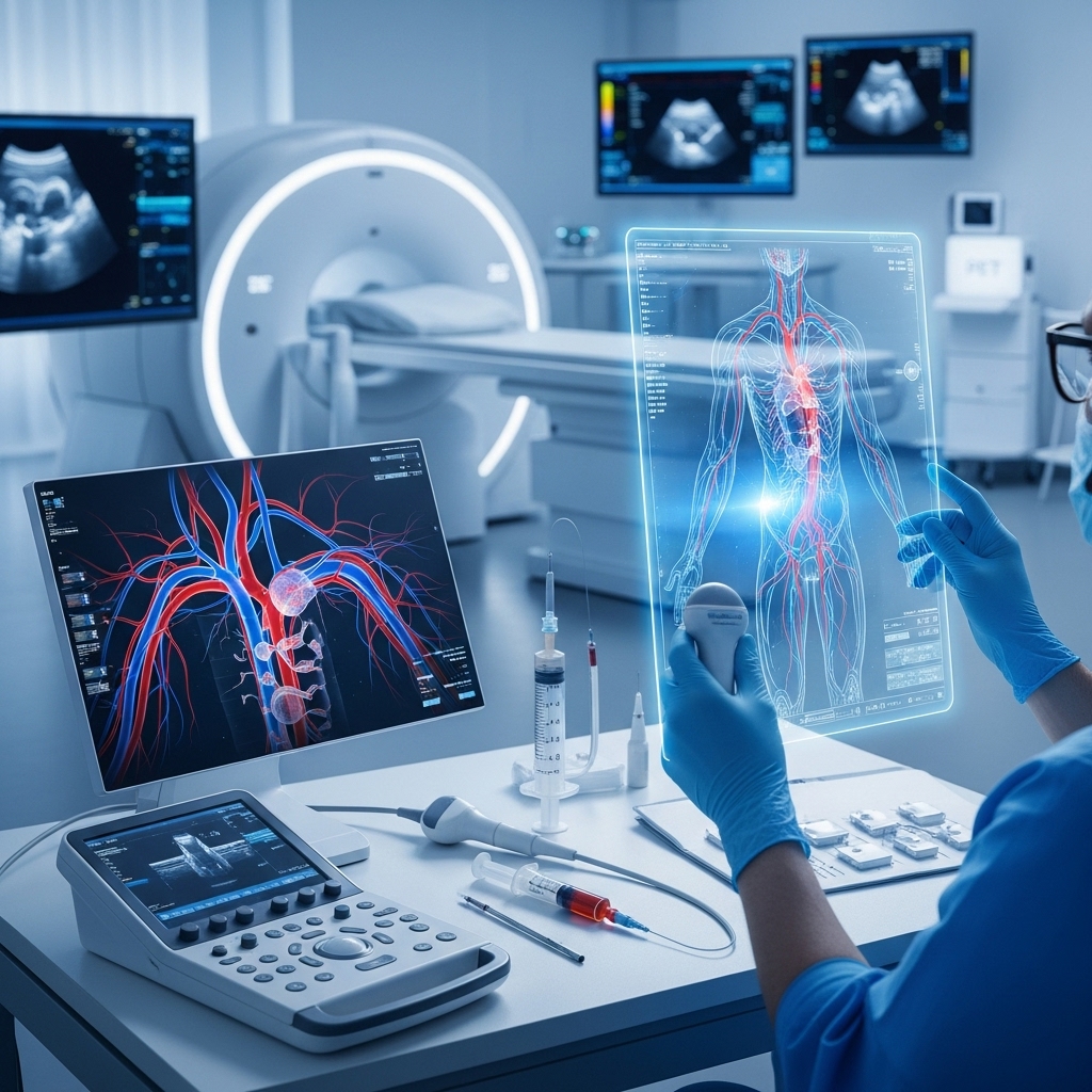
Introduction to Vascular Imaging
Vascular imaging has undergone significant transformations over the years, driven by advances in technology and diagnostic techniques. The field of vascular imaging encompasses a range of modalities aimed at visualizing and assessing the vascular system, which includes arteries, veins, and lymphatic vessels. These advancements have not only improved our understanding of vascular diseases but have also enhanced diagnostic accuracy, leading to better patient outcomes. This article delves into the recent advances in imaging technologies and diagnostic techniques, exploring how they are revolutionizing the field of vascular imaging.
Advances in Imaging Technologies
One of the most significant advancements in vascular imaging is the development of high-resolution imaging technologies. Techniques such as high-frequency ultrasound, magnetic resonance angiography (MRA), and computed tomography angiography (CTA) have become indispensable tools for vascular assessment. For instance, high-frequency ultrasound allows for the detailed examination of superficial vessels, making it ideal for diagnosing conditions like varicose veins or vascular malformations. MRA and CTA, on the other hand, provide comprehensive views of both arterial and venous systems, aiding in the diagnosis of conditions such as atherosclerosis, aneurysms, and deep vein thrombosis.
These imaging technologies have been further enhanced by the integration of contrast agents and advanced software. Contrast-enhanced ultrasound, for example, improves the visualization of blood flow and tissue vascularity, which is particularly useful in assessing tumor vascularization and monitoring response to treatment. Similarly, the development of sophisticated image reconstruction algorithms has significantly improved the resolution and diagnostic capability of CTA and MRA scans.
Diagnostic Techniques in Vascular Imaging
Beyond the advancements in imaging technologies, there have been significant developments in diagnostic techniques used in vascular imaging. One such technique is intravascular ultrasound (IVUS), which involves the use of a small ultrasound probe inserted into the blood vessel to obtain high-resolution images of the vessel wall and lumen. IVUS is particularly useful in coronary interventions, such as stent placement, where it helps in assessing the size of the artery and ensuring proper stent deployment.
Another diagnostic technique that has gained prominence is optical coherence tomography (OCT), which uses low-coherence interferometry to produce high-resolution images of the vascular wall. OCT is advantageous in visualizing the detailed morphology of atherosclerotic plaques and in assessing stent apposition and expansion. These diagnostic techniques, when combined with advanced imaging technologies, offer a powerful toolkit for the comprehensive assessment of vascular diseases.
Applications in Clinical Practice
The advancements in vascular imaging technologies and diagnostic techniques have numerous applications in clinical practice. For instance, in the management of peripheral arterial disease (PAD), CTA and MRA are used to identify and characterize stenotic lesions, guiding endovascular interventions such as angioplasty and stenting. Similarly, in the diagnosis of pulmonary embolism, CTA of the chest has become the gold standard due to its high sensitivity and specificity.
In the realm of neurovascular diseases, imaging plays a critical role in the diagnosis and treatment of conditions such as stroke and aneurysms. Advanced MRI techniques, including diffusion-weighted imaging and perfusion-weighted imaging, are essential in acute stroke management, helping to identify areas of infarction and salvageable tissue. For cerebral aneurysms, 3D angiography provides detailed anatomical information, aiding in the planning of surgical or endovascular treatments.
Future Directions and Challenges
Despite the significant advancements in vascular imaging, there are ongoing challenges and areas for future development. One of the main challenges is the integration of imaging findings with clinical data to provide personalized medicine. The use of artificial intelligence (AI) and machine learning algorithms to analyze large datasets and predict patient outcomes is an area of active research. Additionally, the development of non-invasive and cost-effective imaging modalities that can be used for screening and longitudinal monitoring of vascular diseases is a priority.
Another future direction is the development of molecular imaging techniques that can identify specific biological processes at the molecular level. This could enable the early detection of vascular diseases, even before anatomical changes become apparent. Techniques such as positron emission tomography (PET) with novel tracers are being explored for their potential in imaging atherosclerotic inflammation and vulnerable plaques.
Conclusion
In conclusion, the field of vascular imaging has witnessed tremendous growth, driven by advancements in imaging technologies and diagnostic techniques. These developments have not only enhanced our understanding of vascular diseases but have also improved diagnostic accuracy and patient outcomes. As technology continues to evolve, with the integration of AI, molecular imaging, and personalized medicine, the future of vascular imaging holds much promise. Continued research and innovation are essential to address the current challenges and to further unlock the secrets of the vascular system, ultimately leading to better prevention, diagnosis, and treatment of vascular diseases.
